Peering through MRI scans of fruit and veg
I wrote this article while I was working at Wellcome Collection. It was originally published on their Stacks blog under a CC BY 4.0 license, and is reposted here in accordance with that license.
Having a large image collection to play with means we’ve never short of a good illustration. Recently, somebody used an image of an MRI scan of a pumpkin as the cover picture on an internal blog post. Pretty appropriate in the run-up to Halloween:
It’s one of a series of MRI scans of fruit and vegetables. All the images were created by Alexandr Khrapichev, from the University of Oxford.
An MRI scan is a way to look inside the human body when other types of scanner can’t be used. It’s painless, non-invasive, and can give very high-resolution images. Rather than using x-ray or ultrasound, it uses powerful magnets to affect the movement of water molecules. (Remember that humans are 70% water!) Those movements can be used to construct an image.
Often — as seen here — a single MRI scan takes multiple “slices”. Each slice is a single cross-section of the body, and taking different slices gives you a detailed impression of the body’s interior.
In images like this one, Khrapichev has taken slices of a fruit or vegetable, used colours to match the original, and then stitched them together into a single high-resolution image. It’s a bit of fun, and it nicely illustrates how an MRI scan works.
When I saw these images, I wanted to break them back into individual slices, and combine them into an animated GIF. For me at least, that works a lot better than seeing them laid out on a static page. Since these images are all licensed as CC BY, I’m okay to do that, as long as I link back to the originals!
I’ve been sharing a few examples on my Twitter account, but I wanted to give them a more permanent home. The full resolution images are gorgeous, and the compression on Twitter doesn’t do it justice.
In this post, I’ll show off some of my favourite images. In a sequel, I’ll explain the code I used to create them.
Let’s start with something quite familiar: an apple. Turning it into a GIF shows the shape of the apple, and we can see the gaps in the core:

Here’s another pair of images which are really familiar: a mandarin and a lemon. Within the MRI, you can see the segments of the two citrus fruits — and the occasional flash of a seed.


I love the vibrancy of the bright orange against the black background. The side view is equally impressive:

I’m also rather taken with the red in this tomato. I can see the aspects of a tomato when I look at it, but I don’t think it’s quite as recognisable.

Turning towards the more unusual, this is a kiwifruit. Not only is it a pleasing shade of green, it also shows off the structure nicely. The core and the seeds really stand out in this image:

Going for something even odder, this scan of a passion fruit looks closer to frog spawn than fruit — in fact, it’s the numerous seeds. Once you know what it is, it makes a lot more sense!

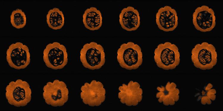
Here’s another one which is quite unusual, and which I find almost mesmerising. It looks like something from the world of microbiology — a cluster of tiny cells — but it’s much larger. Can you guess what it is?

Any ideas? If it helps, here’s another view of the same object, this time from a different angle:

Did you guess correctly?
This is garlic — each segment is an individual clove, which grows as we move from one end towards the centre, then tapers off again at the other end.
There are some even weirder ones. This axial view of a cabbage puts me in mind of the opening credits of Doctor Who. It’s almost hypnotic:

And the other view of the cabbage isn’t any less strange — like a sort of magical forest, the trees growing alongside the path to greet you.

Somehow, these images make cabbage seem much more interesting than when I had it for school dinners.
But what most surely be my favourite bizarre image is the artichoke. The collage image is a bit odd, but stitching them together looks like a portal opening in space. (Has anybody ever used images from an MRI in a science-fiction show?)

And finally, to round out this post, here a GIF of the pumpkin I opened the post with:

Happy Halloween!